Bioprinting Kidney Tumours: A Leap Toward Personalised Cancer Therapy
- Adreeka jeet

- Aug 14, 2025
- 6 min read

A New Frontier in Cancer Modeling
Cancer’s complexity characterised by varying genetics, evolving subclonal populations, and intricate resistance mechanisms has long posed significant hurdles for researchers and clinicians alike. The multifaceted nature of cancer means that no two tumours are exactly alike, which complicates the development of effective treatment strategies. Traditional laboratory models, such as two-dimensional cell cultures and animal models, often fail to accurately capture this heterogeneity, leading to limitations in drug evaluation processes and hindering the translation of research findings into effective patient care. As a result, many promising therapies do not progress beyond clinical trials, and patients may not receive the most effective treatments tailored to their specific tumour profiles. Scientists at Tsinghua University have now tackled this challenge head-on by developing a groundbreaking method to 3D-bioprint kidney tumour organoids utilizing real patient-derived cells. This innovative approach allows for the creation of tumour models that closely mimic the actual architecture and cellular composition of human tumors, achieving a level of fidelity and scalability that was previously out of reach for researchers. By incorporating the unique genetic and phenotypic characteristics of individual patients’ tumours into these organoids, the team has created a more representative platform for drug testing and therapeutic evaluation. The implications of this advancement are profound, as it opens up new avenues for personalized medicine in oncology. With the ability to produce patient-specific tumour organoids, researchers can now conduct more relevant preclinical studies that reflect the complexities of human cancers. This could lead to more accurate predictions of how individual patients will respond to various treatments, ultimately improving the chances of successful outcomes. Furthermore, the scalability of this 3D bioprinting technique means that it could potentially be applied to a wide range of cancer types, paving the way for a more comprehensive understanding of tumour biology and treatment resistance mechanisms across different cancers. In summary, the development of 3D-bioprinted kidney tumour organoids at Tsinghua University represents a significant leap forward in cancer research. By addressing the limitations of traditional lab models and harnessing the power of personalised medicine, this innovative method holds the promise of enhancing drug evaluation processes and improving patient care in the fight against cancer.
Beyond Cells: Recapitulating the Tumour Microenvironment
Rather than printing tumour cells in isolation, the innovative research team at Tsinghua University took a significant step forward by integrating a diverse array of multiple cell types, including endothelial-like cells, into their experimental framework. This strategic inclusion of endothelial-like cells plays a crucial role in the reconstruction of blood vessel structures within the tumours, thereby facilitating a more intricate and realistic model of the tumour microenvironment. By incorporating these various cell types, the team has successfully created a sophisticated model that not only represents the tumour itself but also the surrounding supportive tissues and structures that are essential for tumour growth and development.
This nuanced design allows the resulting organoids to better mimic the complex interactions and dynamics of the tumour microenvironment typically found in vivo. In a natural biological setting, tumours do not exist in isolation; they are embedded within a network of blood vessels, immune cells, and other stromal components that significantly influence their behaviour, growth, and response to therapies. By recreating these conditions, the Tsinghua team enhances the biological realism of their models, which is critical for studying the intricate mechanisms of tumour progression and the efficacy of potential treatments.
Furthermore, this approach not only increases the functional relevance of the organoids but also opens up new avenues for research and development in cancer therapies. It allows scientists to observe how tumours interact with their microenvironment, how they recruit blood vessels for their nutrient supply, and how they evade immune detection. Such insights are invaluable for the development of targeted therapies and for understanding the resistance mechanisms that tumours often employ against conventional treatments. Overall, the integration of multiple cell types into tumour models marks a significant advancement in cancer research, providing a more comprehensive platform for studying the complexities of cancer biology.
Why This Matters: Accuracy, Efficiency, Personalisation:
These lab-grown tumour models known as organoids maintain the unique traits of the original tumour, including structural organisation and cellular behaviors. As a result, they provide a more accurate platform for studying tumor growth, metastasis potential, and drug response patterns. Importantly, the 3D biop
These lab-grown tumour models referred to as organoids are sophisticated biological structures that closely replicate the unique traits of the original tumour. They preserve not only the specific cellular composition of the tumour but also the intricate structural organization and distinct cellular behaviors that characterise the malignancy from which they are derived. This fidelity to the original tumour's architecture and functionality is crucial, as it allows researchers to observe and analyze the tumour's growth dynamics, its potential for metastasis, and the intricate responses to various therapeutic interventions in a controlled environment. As a result, organoids provide a more accurate platform for studying the complexities of cancer biology, enabling scientists to gain insights that are often unattainable through traditional two-dimensional cell cultures or animal models.
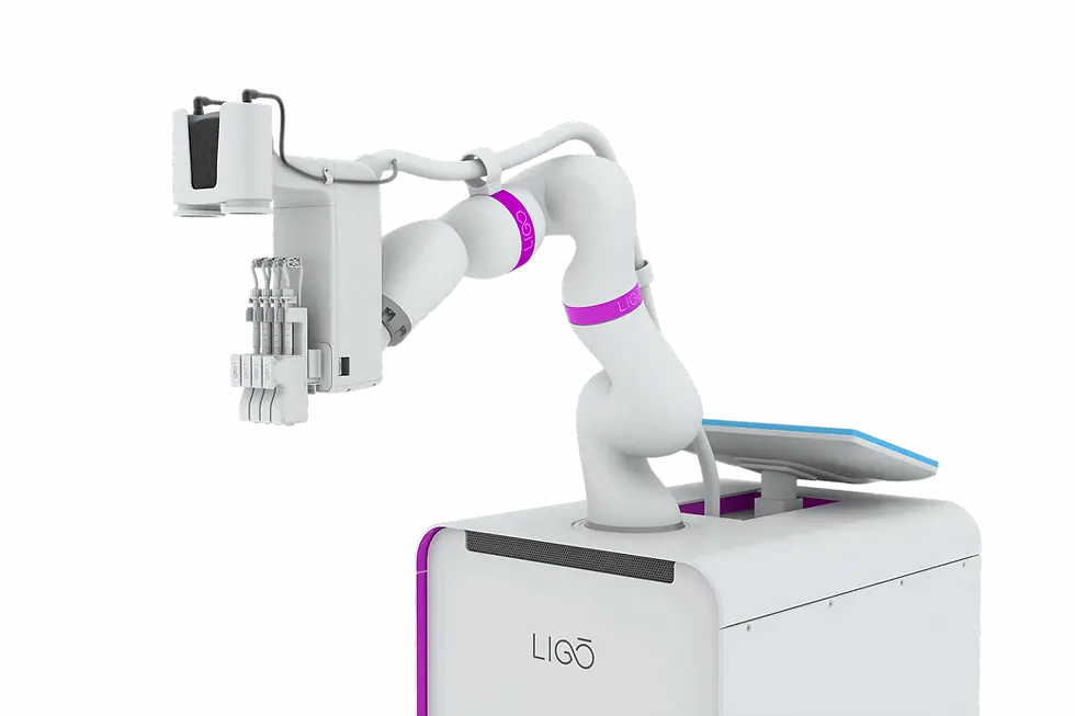
Moreover, the utilization of advanced 3D bioprinting techniques in the creation of these organoids significantly enhances the efficiency and effectiveness of their production. This innovative approach not only streamlines the manufacturing process but also reduces the amount of manual labor required, which can often be time-consuming and prone to variability. By automating the bioprinting process, researchers can produce organoids with remarkable precision and reproducibility, ensuring that each model closely mirrors the characteristics of the original tumor. This capability is particularly beneficial for rapid, scalable testing of multiple therapeutic options, as it allows for the simultaneous evaluation of various drugs and treatment strategies on a wide array of tumor types.
Furthermore, the ability to quickly generate large numbers of organoids opens up new avenues for personalized medicine. By creating organoids from a patient's own tumor cells, clinicians can test the efficacy of different treatments in a laboratory setting before making decisions about the patient's care. This not only enhances the likelihood of selecting the most effective therapy but also minimizes the risk of adverse side effects associated with ineffective treatments. In summary, organoids represent a revolutionary advancement in cancer research and treatment, providing a platform that is not only more representative of actual tumor behavior but also more adaptable to the fast-paced demands of modern medical science.
Bioprinting approach streamlines production, cutting down on manual labor and enabling rapid, scalable testing of multiple therapeutic options.
Toward Precision Medicine in RCC (Renal Cell Carcinoma)
Renal cell carcinoma (RCC) has emerged as a significant global health concern, particularly as its incidence continues to rise in various populations worldwide. This type of cancer originates in the kidneys and is known for its aggressive nature and complex biological behaviour. The management of RCC presents numerous challenges, primarily due to the high variability in how individual patients respond to different treatment modalities. This variability can be attributed to a multitude of factors, including genetic differences, tumour heterogeneity, and the unique microenvironment of each tumor. Moreover, the phenomenon of widespread drug resistance complicates treatment efforts, as many patients may initially respond to therapy but later experience disease progression due to the cancer's ability to adapt and evade therapeutic agents. This resistance can manifest through various mechanisms, including mutations in the cancer cells or alterations in drug metabolism. Additionally, the frequent recurrence of RCC after initial treatment poses another significant hurdle for healthcare providers and patients alike, leading to a cycle of ongoing surveillance and repeated interventions. In response to these challenges, the development of patient-specific tumor models has emerged as a promising strategy in the field of oncology. These models, which are created by taking cells from a patient’s tumor and cultivating them in a laboratory setting, allow researchers and clinicians to study the tumor's characteristics in a controlled environment. By analyzing these models, it becomes possible to gain insights into the tumor's behavior, its response to various treatments, and the underlying mechanisms driving resistance. This innovative approach enables more informed personalized treatment selection, as clinicians can tailor therapeutic strategies based on the specific characteristics and responses of an individual patient's tumor. Rather than relying on a one-size-fits-all approach, which often involves a trial-and-error process that can be both time-consuming and emotionally taxing for patients, personalized treatment plans can be developed that are more likely to be effective. This not only has the potential to improve patient outcomes but also to enhance the overall efficiency of cancer treatment, reducing unnecessary exposure to ineffective therapies and their associated side effects. In summary, as RCC continues to pose a significant challenge in the realm of cancer care, the integration of patient-specific tumor models into clinical practice represents a significant advancement. This method not only addresses the complexities associated with treatment variability and drug resistance but also paves the way for a new era of personalized medicine in oncology, where treatment decisions are informed by the unique biological characteristics of each patient’s tumor.
Scaling Up: Turning Lab Innovation into Clinical Impact
Dr. Yuan Pang, a co-author of the study, underscores the shift from experimentation to application: “This new method could greatly improve how we study kidney cancer and develop personalized treatments for patients. The rapid production of organoids will make it much faster to find the right treatment for individual patients"

Tsinghua University


Comments